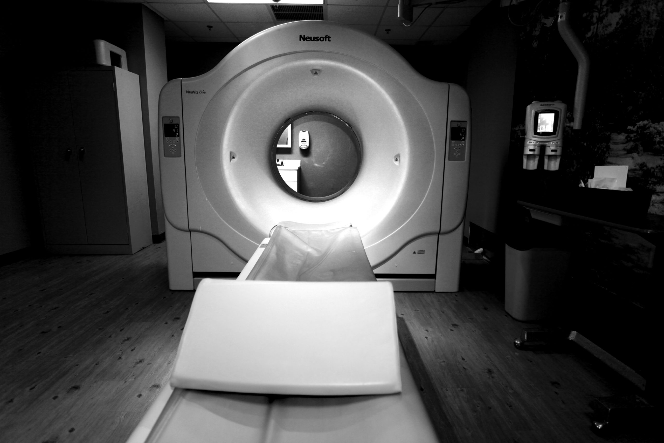
CT

- 64 Multi-Slice capability offering diagnostic advantages such as
- Better quality
- New applications with 3-D volume reconstruction
- More comprehensive diagnosis in less time
- High speed scanning offers patients
- Shorter exam times
- Single breath-hold scanning
- Rapid results
- Metal Artifact Reduction
CT (also called computed tomography or a CAT scanner) has become one of the most important tools to diagnoses head and spine injuries, lung and liver disease, cancer, tumors, blood clots, internal bleeding, and other diseases and illnesses. CT exams are especially useful in the emergency department, where faster diagnosis can be lifesaving for trauma cases like auto accident victims.
The 64-Slice CT can produce 3-D pictures of anatomical structures like aneurysms, tumors, and infections. When physicians can see what’s inside better, they make more accurate diagnoses.
During a CT exam, a patient lies on a table and is slowly moved into the large donut-shaped opening of the scanner. Once inside, a series of X-ray beams create hundreds of cross-sectional pictures that represent slices of the patient’s body. Seconds later, the system’s computer assembles the slices into images that are interpreted by a radiologist.
The 64-Slice CT Scanner can acquire more of those anatomical slices, thanks to a new technology called multi-slice imaging.
As a result, the CT’s multi-slice technology is quick enough to capture images of the body’s organs. Multi-slice imaging is also especially useful for examining patients who are unable to hold their breath, like trauma victims, acutely ill patients and young children.
To learn more about CT, click here

